| Home | E-Submission | Sitemap | Editorial Office |  
J Minim Invasive Spine Surg Tech > Volume 9(Suppl 2); 2024 > Article
Hammed, Mahfoud, Alkhanous, Abboud, Qasem, and Hasan: Management of Spinal Myeloid Sarcoma in a 23-Year-Old Woman With a History of Acute Myeloid Leukemia

Abstract

Myeloid sarcoma (MS) is an uncommon extramedullary tumor comprising immature myeloid cells, primarily associated with acute myeloid leukemia (AML). Spinal involvement is observed in fewer than 1% of cases. We present an unusual case of extramedullary MS in a patient with AML. A 23-year-old woman with a history of AML presented with neurological symptoms, including limb pain, weakness, and numbness. Imaging revealed an intramedullary enhancing mass at the cervical level. Partial laminectomy and microsurgical resection were performed, followed by laminoplasty. Immunohistochemistry supported the diagnosis, confirming MS. The patient was subsequently transferred to the hematologic department and received tailored treatment. The diverse clinical manifestations of spinal MS pose diagnostic challenges. In this case, an atypical intramedullary location led to incorrect initial diagnostic impressions. Comprehensive evaluations, including repeated biopsies and immunohistochemistry, were crucial for an accurate diagnosis. Treatment strategies vary based on systemic involvement, and prompt diagnosis is important for favorable outcomes. MS, especially in the spine, presents diagnostic and therapeutic complexities. Timely recognition, an accurate diagnosis through comprehensive evaluations, and tailored treatment strategies are crucial for positive outcomes in this rare hematologic entity.

INTRODUCTION

Myeloid sarcoma, or MS, is a rare extramedullary tumor consisting of immature myeloid cells [1]. It can manifest in patients with acute myeloid leukemia (AML), myelodysplastic syndrome, or chronic myelogenous leukemia. The incidence of MS in AML patients ranges from 2% to 10.4%, with most cases associated with AML FAB M2, M4, and M5 [2,3]. Certain cytogenetic abnormalities in AML, such as t(8;21), have been linked to a higher incidence of MS [4]. The overall prevalence of MS in any organ is 2.9% among all patients with acute and chronic myeloid leukemia (CML). Specifically, the prevalence of MS involving the spine is 1.0% among these patients [5,6]. We report an extremely rare case of intradural extramedullary MS n a patient with AML as a hematologic relapse.

CASE REPORT

In this case, a 23-year-old female presented with left upper limb pain, weakness in the left hemimuscles, numbness in all 4 limbs, and difficulty walking. The initial manifestation as cervical pain progressed to radicular pain of the sixth cervical nerve, resulting in weakness (4 out of 5 strength in most muscles) and numbness in the left upper limb. Additionally, weakness (4 out of 5 strength) was noted in various muscle groups of the left lower limb. Reflex examinations indicated hyperactive tendon reflexes, a negative Hoffman's sign, and a positive Babinski's reflex.
Of significance, the patient's medical history revealed a diagnosis of AML from 2019 to 2022, highlighting a pertinent health background. Furthermore, the patient had a known sensitivity to soybeans.
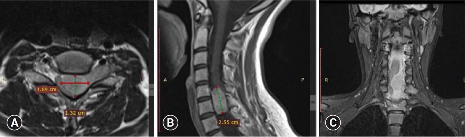
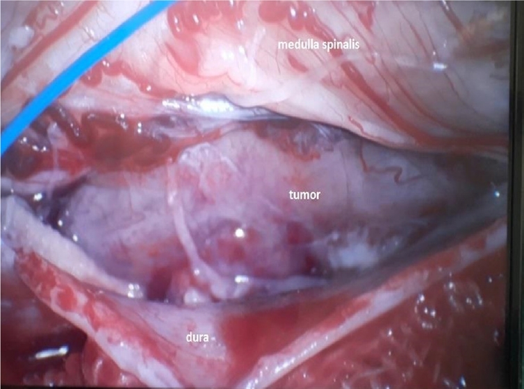
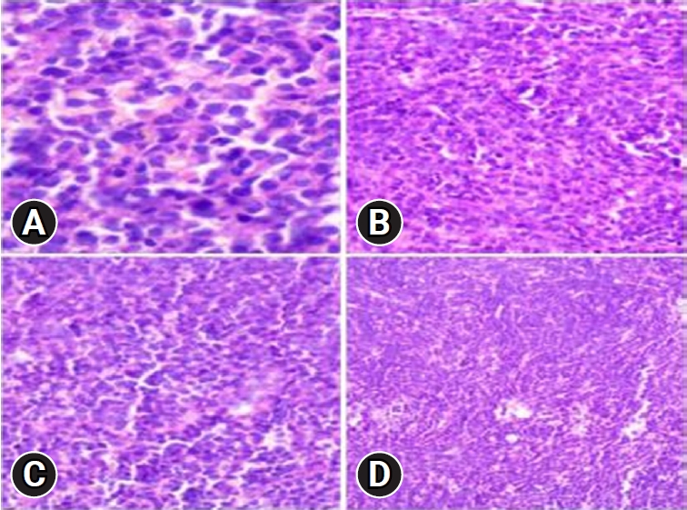
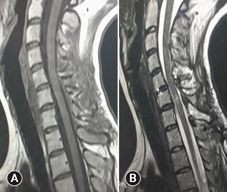
Upon admission, an enhanced spinal magnetic resonance imaging (MRI) revealed an intramedullary enhancing mass lesion, along with changes at the C5–7 level (Figure 1). The initial impression led to suspicions of granulocytic sarcoma or atypical infectious myelitis, prompting a consultation with the neurosurgery department for an open biopsy. The intervention commenced with a posterior midline incision, strategically chosen to provide optimal access to the cervical spine. The subsequent step involved a precise partial cut of the lamina, a critical maneuver to facilitate further exploration. The surgical field was then meticulously refined, as the dura mater, the protective covering of the spinal cord, was opened with utmost precision under the microscope. Following this delicate stage, tumor resection ensued with meticulous care, with a primary focus on minimizing damage to surrounding structures, particularly the nearby nerves. The surgical team executed laminoplasty using small titanium plates, securing them in place with screws to ensure stability and structural support. The spine, identified as intradural extramedullary with adhesion to spinal roots (Figure 2), was successfully resected without complications, surgical resection aspects were piecemeal with histological margins type R1, and followed by laminoplasty. After the operation, numbness and pain in the 4 limbs decreased, with improvement in pain in the left upper limb and 3 months after surgery the patient’s neurological examination was normal. Frozen biopsy results revealed malignancy with atypical small round cells (Figure 3).
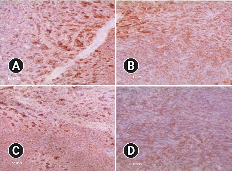
Final pathologic confirmation diagnosed the condition as MS, supported by positive immunohistochemistry for myeloperoxidase (MPO), leukocyte common antigen (LCA), and vimentin, and negativity for creatine kinase, desmin, synaptophysin, and CD99. Histologic findings indicated numerous blasts with round or oval nuclei, dispersed chromatin, prominent nucleoli, and agranular cytoplasm with varying basophilia. Eosinophilic granulation was notable in a minority of cell populations (Figure 4). Following histologic confirmation, the patient was transferred to the oncology department, and she underwent supplementary radiotherapy after surgery for 2 months and is currently under follow-up (Figure 5).
This case underscores the complexity of neurological symptoms in individuals with a history of leukemia and emphasizes the crucial role of comprehensive diagnostic procedures in achieving an accurate diagnosis for appropriate therapeutic interventions.
Written informed consent was obtained from the patient for publication of this case report and accompanying images.

DISCUSSION

MS, initially described in 1811, represents a rare hematologic phenomenon occurring in patients with myeloid leukemia [7,8]. Its prevalence in the spine is estimated to be less than 1% among all patients with acute and CML. While it can affect any level of the spine, it is more frequently observed in the lumbosacral spine, followed by the thoracic and cervical levels [9-11]. Clinical manifestations of spinal MS vary, depending on the tumor location and size [12-15]. Typically, spinal MS in the epidural or paraspinal tissues presents with signs of cord compression, resulting from a mass effect [14,16,17]. However, in some cases, as observed in the presented case, atypical intradural extramedullary location can lead to an unspecific presentation, potentially misdiagnosed as conditions like acute transverse myelitis or demyelinating diseases, leading to delayed diagnosis [8,9,13,18].
The diagnosis of spinal MS poses challenges as clinical and laboratory findings are often non-contributory [13,19]. Radiologic findings are usually non-specific, with the mass appearing as a soft tissue mass [16]. On MRI examination, spinal MS exhibits similar signal intensity to muscle on T1-weighted images and intermediate signal intensity on T2-weighted images, with subtle to intermediate enhancement with gadolinium [12,15,20]. Immunohistochemistry emerges as the most accurate method for confirming the diagnosis of MS, utilizing a series of markers for diagnosis confirmation and further lineage characterization [10,14,18,21].
MS can occur in patients without evidence of leukemia and may precede the development of systemic disease [13,14]. In cases where leukemia is established, the diagnosis of MS is relatively straightforward and should be considered in the differential diagnosis for patients with leukemia presenting with a soft tissue mass [21,22]. However, in the absence of a clinical history of leukemia, obtaining a pathologic diagnosis becomes crucial, as illustrated in the presented case, where repeated open biopsy and immunohistochemical stains aided in confirming the diagnosis.
The treatment strategy for MS hinges on systemic manifestations of leukemic cells and whether it represents the initial diagnosis or relapse [10,11,21]. The optimal timing and approach to isolated MS remain controversial. Delayed or inadequate systemic treatment often progresses to systemic infiltrations [15,19,21,23]. For patients with MS and concurrent marrow involvement, systemic treatment is recommended [13,14,18]. Prompt diagnosis and adequate systemic treatment are essential for a favorable outcome.
The differential diagnosis of MS in the head and neck region encompasses various malignant lymphomas, blastic plasmacytoid dendritic cell neoplasm, solid tumors (carcinoma and melanoma), and small blue cell tumors of childhood. It is crucial to distinguish MS from other entities, such as diffuse large B-cell lymphoma, Burkitt lymphoma, and lymphoblastic lymphoma/leukemia, emphasizing the significance of histological, flow cytometric, and cytogenetic analyses for accurate diagnosis.

CONCLUSION

MS, particularly in the spine, presents unique challenges in diagnosis and treatment. Timely recognition, accurate diagnosis through comprehensive evaluations, and tailored treatment strategies, considering the systemic manifestations and risk factors, are critical for achieving positive outcomes in patients with this rare hematologic phenomenon.

NOTES

Conflict of Interest

The authors have nothing to disclose.

Funding/Support

This study received no specific grant from any funding agency in the public, commercial, or not-for-profit sectors.

Figure 1.
Enhanced spinal magnetic resonance imaging showing an intramedullary enhancing mass lesion, along with changes at the C5–7 level. Axial (A), sagittal (B), and coronal (C).
jmisst-2023-01144f1.jpg
Figure 2.
Intraoperative image showing an intradural extramedullary tumor with adhesion to the spinal root.
jmisst-2023-01144f2.jpg
Figure 3.
Frozen biopsy demonstrating a malignancy with atypical small round cells (hematoxylin and eosin stain, x400 [A], x200 [B], x200 [C], x100 [D]).
jmisst-2023-01144f3.jpg
Figure 4.
Immunohistochemistry showing positive immunohistochemistry for myeloperoxidase (MPO; A, x40), leukocyte common antigen (LCA; B, x40), vimentin (C, x40), and negativity for creatine kinase, desmin, synaptophysin, and CD99 (D, x40).
jmisst-2023-01144f4.jpg
Figure 5.
Postoperative T1-weighted (A) and T2-weighted sagittal magnetic resonance imagings (B) showing full removal of the tumor.
jmisst-2023-01144f5.jpg

REFERENCES

1. Bakst RL, Tallman MS, Douer D, Yahalom J. How I treat extramedullary acute myeloid leukemia. Blood 2011;118:3785–93.
crossref pmid pdf
2. van der Weide M, Imandt LM, Langenhuijsen MM. Motility of leukaemic cells in acute leukaemia related to tumour mass, maturation and FAB classification. Acta Haematol 1988;80:34–9.
crossref pmid pdf
3. Shimizu H, Saitoh T, Hatsumi N, Takada S, Yokohama A, Handa H, et al. Clinical significance of granulocytic sarcoma in adult patients with acute myeloid leukemia. Cancer Sci 2012;103:1513–7.
crossref pmid pmc
4. Tallman MS, Hakimian D, Shaw JM, Lissner GS, Russell EJ, Variakojis D. Granulocytic sarcoma is associated with the 8;21 translocation in acute myeloid leukemia. J Clin Oncol 1993;11:690–7.
crossref pmid
5. Novick SL, Nicol TL, Fishman EK. Granulocytic sarcoma (chloroma) of the sacrum: initial manifestation of leukemia. Skeletal Radiol 1998;27:112–4.
crossref pmid pdf
6. Seok JH, Park J, Kim SK, Choi JE, Kim CC. Granulocytic sarcoma of the spine: MRI and clinical review. AJR Am J Roentgenol 2010;194:485–9.
crossref pmid
7. Devi VL, Doval DC, Rao CR, Bapsy PP. Spinal epidural granulocytic sarcoma: a rare presentation of acute myeloid leukemia. Ann Saudi Med 1996;16:212–4.
pmid
8. Struhal W, Oberndorfer S, Lahrmann H, Lindeck-Pozza E, Hess B, Nussgruber V, et al. Myeloid sarcoma in the central nervous system: case report and review of the literature. Acta Clin Croat 2008;47:19–24.
pmid
9. Kyaw TZ, Maniam JA, Bee PC, Chin EF, Nadarajan VS, Shanmugam H, et al. Myeloid sarcoma: an unusual presentation of acute promyelocytic leukemia causing spinal cord compression. Turk J Haematol 2012;29:278–82.
crossref pmid pmc
10. Yang B, Yang C, Fang J, Yang J, Xu Y. Clinicoradiological characteristics, management and prognosis of primary myeloid sarcoma of the central nervous system: a report of four cases. Oncol Lett 2017;14:3825–31.
crossref pmid pmc
11. Yang C, Liu Y, Li G, Bai J, Qian J, Xu Y. Multifocal myeloid sarcoma in the central nervous system without leukemia. Clin Neurol Neurosurg 2014;120:99–102.
crossref pmid
12. Eom KS, Kim TY. Intraparenchymal myeloid sarcoma and subsequent spinal myeloid sarcoma for acute myeloblastic leukemia. J Korean Neurosurg Soc 2011;49:171–4.
crossref pmid pmc
13. Hu X, Shahab I, Lieberman IH. spinal myeloid sarcoma "chloroma" presenting as cervical radiculopathy: case report. Global Spine J 2015;5:241–6.
crossref pmid pmc pdf
14. Olar A, Lapadat R, Davidson CJ, Stein TD, Dahiya S, Perry A, et al. Central nervous system involvement by myeloid sarcoma: a report of 12 cases and review of the literature. Clin Neuropathol 2016;35:314–25.
crossref pmid
15. Widhalm G, Dietrich W, Müllauer L, Streubel B, Rabitsch W, Kotter MR, et al. Myeloid sarcoma with multiple lesions of the central nervous system in a patient without leukemia. Case report. J Neurosurg 2006;105:916–9.
pmid
16. Cho SF, Liu TC, Chang CS. Isolated central nervous system relapse presenting as myeloid sarcoma of acute myeloid leukemia after allogeneic peripheral blood stem cell transplantation. Ann Hematol 2013;92:133–5.
crossref pmid pdf
17. Stepensky P, Revel-Vilk S, Yehuda-Gafni O, Mali B, Resnick IB, Weintraub M. Isolated central nervous system granulocytic sarcoma and meningeal myeloid leukemia: successful treatment without radiotherapy. Isr Med Assoc J 2009;11:569–70.
pmid
18. Yang C, Fang J, Xu Y. Isolated spinal myeloid sarcoma with rapid progression. Spine J 2016;16:e517–8.
crossref pmid
19. Serefhanoglu S, Goker H, Aksu S, Buyukasik Y, Sayinalp N, Haznedaroglu IC, et al. Spinal myeloid sarcoma in two non-leukemic patients. Intern Med 2010;49:2493–7.
crossref pmid
20. Best-Aguilera CR, Vazquez-Del Mercado M, Muñoz-Valle JF, Herrera-Zarate L, Navarro-Hernandez RE, Martin-Marquez BT, et al. Massive myeloid sarcoma affecting the central nervous system, mediastinum, retroperitoneum, liver, and rectum associated with acute myeloblastic leukaemia: a case report. J Clin Pathol 2005;58:325–7.
crossref pmid pmc
21. Isshiki Y, Ohwada C, Togasaki E, Shimizu R, Hasegawa N, Yamazaki A, et al. Acute myeloid leukemia concurrent with spinal epidural extramedullary myeloid sarcoma accompanied by a high CD25 expression and the FLT3-ITD mutation. Intern Med 2014;53:1159–64.
crossref pmid
22. Takeda M, Yamaguchi S, Eguchi K, Kajiume T, Nishimura S, Kobayashi M, et al. Spinal epidural granulocytic sarcoma in a child precedent to clinical manifestation of acute myeloid lymphoma: case report. Neurol Med Chir (Tokyo) 2009;49:221–4.
crossref pmid
23. Landis DM, Aboulafia DM. Granulocytic sarcoma: an unusual complication of aleukemic myeloid leukemia causing spinal cord compression. A case report and literature review. Leuk Lymphoma 2003;44:1753–60.
crossref pmid
TOOLS
PDF Links  PDF Links
PubReader  PubReader
ePub Link  ePub Link
XML Download  XML Download
Full text via DOI  Full text via DOI
Download Citation  Download Citation
  Print
Share:      
METRICS
0
Crossref
0
Scopus
342
View
4
Download
Related article
About |  Browse Articles |  Editorial Policy |  For Contributors
Editorial Office
Department of Neurosurgery, Harrison Spinartus Hospital Chungdam
646 Samseong-ro, Gangnam-gu, Seoul 06084, Korea
TEL: +82-2-6003-9767    FAX: +82-2-3445-9755   E-mail: office@jmisst.org
Publisher
Korean Minimally Invasive Spine Surgery Society
350 Seocho-daero, Seocho-gu, Seoul 06631, Korea
TEL: +82-2-585-5455    FAX: +82-2-523-6812   E-mail: komisskomiss@gmail.com
Copyright © Korean Minimally Invasive Spine Surgery Society.                 Developed in M2PI